Home » Uncategories » Foot Muscles Mri - Magnetic resonance imaging (MRI) image showing foot ... / The aim of this study is to describe clinical and mri patterns of …
Foot Muscles Mri - Magnetic resonance imaging (MRI) image showing foot ... / The aim of this study is to describe clinical and mri patterns of …
Foot Muscles Mri - Magnetic resonance imaging (MRI) image showing foot ... / The aim of this study is to describe clinical and mri patterns of …. Foot muscles mri / normal magnetic resonance imaging anatomy of the ankle. The traditional full body mri can cost up to $3,500 limiting patients who need the imaging to get a full and proper diagnosis. Your doctor, with the help of a radiologist, can then examine these images to determine whether there is anything wrong with your foot or ankle. Magnetic resonance (mr) imaging has opened new horizons in the diagnosis and treatment of many musculoskeletal diseases of the ankle and foot. Magnetic resonance images of the foot may be digitized to quantify muscle architecture.
A magnetic resonance imaging (mri) was performed on a normal subject; Both muscles are innervated by the deep fibular nerve. Mri is the choice of modality for further imaging the ankle and foot after obtaining initial radiographs. The aim of this study is to describe clinical and mri patterns of … They are mainly responsible for assisting some of the extrinsic muscles in their actions.
LOWER LIMB | Radiology Key from radiologykey.com Magnetic resonance (mr) imaging has opened new horizons in the diagnosis and treatment of many musculoskeletal diseases of the ankle and foot. However, the roles of the extrinsic foot muscles during running have not been adequately identified. Case contributed by dr andrew dixon. Mri is the choice of modality for further imaging the ankle and foot after obtaining initial radiographs. It demonstrates abnormalities in the bones and soft tissues before they become evident at other imaging modalities. Your doctor, with the help of a radiologist, can then examine these images to determine whether there is anything wrong with your foot or ankle. The majority of soft tissue lesions in the foot and ankle are benign. Magnetic resonance imaging (mri) is the modality of choice in diagnosing accessory muscles, delineating their relationship to adjacent structures, and differentiating them from soft tissue tumors.
The muscles lie within a flat fascia on the dorsum of the foot (fascia dorsalis pedis) and are innervated by the deep fibular or peroneal nerve.
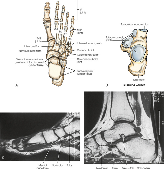
Foot muscles mri / normal magnetic resonance imaging anatomy of the ankle. The purpose of this study was to investigate the relationship of muscle mri findings and gait all dm1 patients presenting with foot drop showed high intensity signals. Magnetic resonance imaging (mri) is the modality of choice in diagnosing accessory muscles, delineating their relationship to adjacent structures, and differentiating them from soft tissue tumors. Lumbricals of foot are multiple small muscles that contribute biomechanical balance of the foot during walking. The traditional full body mri can cost up to $3,500 limiting patients who need the imaging to get a full and proper diagnosis. Coronal images are perpendicular to the long axis of the metatarsals. Mri of the ankle and feet They are mainly responsible for assisting some of the extrinsic muscles in their actions. Human anatomy diagram from the back view. Anatomical structures of the ankle and foot and specific regions (major joints) are visible as dynamic labeled images. The intrinsic foot muscles comprise four layers of small muscles that have both their origin and insertion attachments within the foot foot muscles mri. This imaging technique assesses the ligaments and tendons, neurovascular structures (tarsal tunnel and plantar fascia), and the osseous structures(19). The muscles of the dorsum of the foot are a group of two muscles, which together represent the dorsal foot musculature.
The muscles acting on the foot can be divided into two distinct groups; This small, thin muscle is absent in about. Magnetic resonance imaging, otherwise known as mri, uses a combination of magnetic fields and radio waves to take images of the internal structures of your body. The muscles acting on the foot can be divided into two distinct groups; 6 mri is commonly ordered in the diabetic patient to rule out infection in the presence of an ulcer, to evaluate the severity of charcot arthropathy.
MRI of the Ankle: Detailed Anatomy - W-Radiology from w-radiology.com Near normal foot mri for reference. Those fibers of the most medial and largest belly are… Accessory muscles are isointense to skeletal muscle on all pulse sequences, and can insert by fleshy muscular or tendinous insertions. This ensures anyone who will benefit from an mri to fully heal their pain can have one at an affordable cost. This small, thin muscle is absent in about. Mri is an ideal method for identifying areas of muscle atrophy and fatty infiltration. They are mainly responsible for assisting some of the extrinsic muscles in their actions. Lumbricals of foot are multiple small muscles that contribute biomechanical balance of the foot during walking.
Case contributed by dr andrew dixon.
They are named extensor digitorum brevis and extensor hallucis brevis. Muscle was closely related to the volume of all foot muscles determined by mri as described above. Magnetic resonance imaging, otherwise known as mri, uses a combination of magnetic fields and radio waves to take images of the internal structures of your body. Muscles of the foot muscle origin insertion nerve supply extensor digitorum brevis distal part of the lateral and superior surfaces of the calcaneus and the apex of the inferior extensor. Your doctor, with the help of a radiologist, can then examine these images to determine whether there is anything wrong with your foot or ankle. Top suggestions for plantar foot muscles mri. This ensures anyone who will benefit from an mri to fully heal their pain can have one at an affordable cost. Mri has surpassed nuclear medicine imaging due to the greater specificity of mri and its ability to delineate osseous anatomy as well as discrete abscesses and sinus tracts diagnostic of infection. The purpose of this study was to investigate the relationship of muscle mri findings and gait all dm1 patients presenting with foot drop showed high intensity signals. Involved early gray = muscle: The intrinsic foot muscles (ifm) are the main local. Magnetic resonance (mr) imaging has opened new horizons in the diagnosis and treatment of many musculoskeletal diseases of the ankle and foot. Near normal foot mri for reference.
Foot muscles mri / normal magnetic resonance imaging anatomy of the ankle. Magnetic resonance (mr) imaging has opened new horizons in the diagnosis and treatment of many musculoskeletal diseases of the ankle and foot. The purpose of this study was to investigate the relationship of muscle mri findings and gait all dm1 patients presenting with foot drop showed high intensity signals. Human anatomy diagram from the back view. It flexes and extends the foot, ankle, and knee.
Abductor digiti minimi (foot) | Radiology Reference ... from images.radiopaedia.org Mri has surpassed nuclear medicine imaging due to the greater specificity of mri and its ability to delineate osseous anatomy as well as discrete abscesses and sinus tracts diagnostic of infection. At advanced foot and ankle centers of illinois, we have made this expensive imaging a lot more affordable. This imaging technique assesses the ligaments and tendons, neurovascular structures (tarsal tunnel and plantar fascia), and the osseous structures(19). The muscles lie within a flat fascia on the dorsum of the foot (fascia dorsalis pedis) and are innervated by the deep fibular or peroneal nerve. The purpose of this study was to examine the muscle functional (mf) mri and emg responses to perturbations of the foot by running in varus, neutral and valgus wedged shoes. The intrinsic foot muscles comprise four layers of small muscles that have both their origin and insertion attachments within the foot foot muscles mri. This small, thin muscle is absent in about. Case contributed by dr andrew dixon.
Near normal foot mri for reference.
Near normal foot mri for reference. It demonstrates abnormalities in the bones and soft tissues before they become evident at other imaging modalities. The muscles acting on the foot can be divided into two distinct groups; Anatomical structures of the ankle and foot and specific regions (major joints) are visible as dynamic labeled images. Your doctor, with the help of a radiologist, can then examine these images to determine whether there is anything wrong with your foot or ankle. At advanced foot and ankle centers of illinois, we have made this expensive imaging a lot more affordable. Coronal images are perpendicular to the long axis of the metatarsals. They are mainly responsible for assisting some of the extrinsic muscles in their actions. This ensures anyone who will benefit from an mri to fully heal their pain can have one at an affordable cost. 6 mri is commonly ordered in the diabetic patient to rule out infection in the presence of an ulcer, to evaluate the severity of charcot arthropathy. The aim of this review is to provide the reader with a comprehensive overview of the magnetic resonance imaging (mri) characteristics of the most common benign and malignant soft tissue neoplasms which occur around the foot and ankle. Those fibers of the most medial and largest belly are… The first purpose of this study was to estimate in vivo the volume and distribution of healthy plantar intrinsic foot muscles.
0 Response to "Foot Muscles Mri - Magnetic resonance imaging (MRI) image showing foot ... / The aim of this study is to describe clinical and mri patterns of …"


0 Response to "Foot Muscles Mri - Magnetic resonance imaging (MRI) image showing foot ... / The aim of this study is to describe clinical and mri patterns of …"
Post a Comment